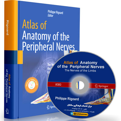
Atlas of Anatomy of the Peripheral Nerves – The Nerves of the Limbs

مولف

Daniel Thomas Ginat

ناشر

‎ Springer

ISBN

978-3030491819

سال انتشار

2020

قطع

رحلی A4 استاندارد

تعداد صفحات

504

تخصص

Anatomy, Neurology, Neurosurgery, Plastic Surgery, Anesthesia

ویراست

1st Edition

تعداد DVD فیلم

1

👏👏👈 در این کتاب شما می تونید کاملترین آناتومی اعصاب محیطی رو مشاهده کنید
❤️در ابتدا تمام اعصاب محیطی به تفکیک اندام فوقانی و اندام تحتانی از هم جدا می گردد
☀️👈 سپس هر عصب به صورت ریز و موشکافانه ای تحت بررسی قرار گرفته و آناتومی آن در کاملترین شکل ممکن به تصویر کشیده شده است

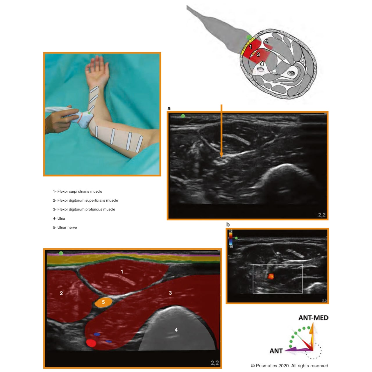
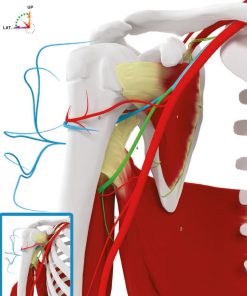
این آناتومی شامل آناتومی شماتیک است که در آن 🔸 منشا عصب 🔸 مسیر حرکت 🔸 انشعابات عصب 🔸 عضلات مرتبط با عصب به خوبی نشان داده شده است.

🔔💥 پس از این به سراغ آناتومی سونوگرافی عصب مربوطه می رود :

بهترین پوزیشن بیمار و نحوه گرفتن و مسیر حرکت پروب در سونوگرافی هر عصب نشان داده شده است.
و همچنین در هر عصب راهنمایی هایی هم جهت انجام تزریقات و جراحی آن عصب بیان شده است.

🎥📀 و اما در مورد مجموعه فیلمهای کتاب که بسیار ارزشمند و کاربردی است در کنار فیلم 3 بعدی که عصب و عضله و نحوه حرکت پروب سونوگرافی روی عضله را نشان میدهد در سمت مقابل نیز فیلم سونوگرافی که اجزاء آن به تفکیک مشخص شده است نشان داده می شود

This book focuses on the anatomy of the peripheral nervous system. Using the latest 3D-computer graphic modeling techniques, the author developed the innovative NEURO 3D LOCATORTM concept, which provides 3D in-vivo ultrasound images of peripheral nerve architectures, allowing readers to develop a mental real-time 3D GPS of the peripheral nervous system.  This new edition is an extended version of the “Student edition” dedicated to Experts and is divided into three main parts: The first part describes fundamental concepts, from immunohistochemistry to limb innervation, and includes a detailed evaluation of the morphofunctional anatomy of the peripheral nerves. It also presents relevant data on neuromuscular transmission, from both classic and recent literature, to enable readers to gain an understanding the physiology and pathology of peripheral nerves as well as the prospects of repair. The second section addresses the upper limb, the brachial plexus and related peripheral nerves, while the third section focuses on the lower limb, the lumbosacral plexus and related peripheral nerves. By providing MRI sections related to the drawings and the descriptions of main nerve injuries, it facilitates radiological interpretation and clinical learning. The book also features detailed descriptions of surgical approaches and the ultrasound anatomy of the limbs, and includes supplementary material on applications to peripheral nerve stimulation, surgical procedures and interventional pain medicine techniques. Presenting high-quality 3D videos showing the progression of the ultrasound probe in real-time, synchronized with live ultrasound views and enhanced with anatomical computerized graphic layers, as well as over 500 outstanding full-color 2D and 3D illustrations, and access to than 100 practical videos, this unique book is a valuable resource for anesthesiologists, radiologists, orthopedic surgeons, neurosurgeons, neuromodulators, physiatrists, pain physicians and rheumatologists. It will also appeal to the medical community in general.

جهت مشاهده قیمت و خرید محصول پس از انتخاب نوع صحافی دلخواه و تعداد مورد نظر ، محصول را به سبد خرید اضافه کنید
پاک کردن

Total

▼ محصولات مرتبط ▼

▼ نمونه صفحات ▼

نمونه صفحات