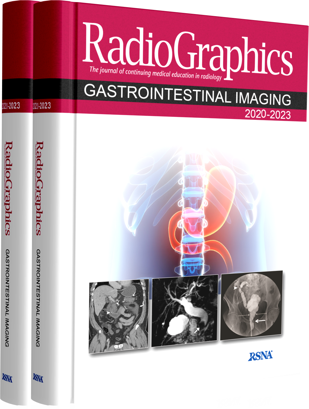
RadioGraphics یک مجله ادواری است که تحت نظارت هیئت مدیره انجمن رادیولوژی آمریکا، شرکت (RSNA) منتشر می شود، انجمن رادیولوژی آمریکا بر ماهیت همه مطالب انتخاب شده برای انتشار، نظارت می کند.
نظر به گستره وسیع اطلاعات در حوضه رادیولوژی و تخصصی شدن مطالعه و فعالیت رادیولوزیست ها در مراکز رادیولوژی قسمت های مختلف این مجلات به صورت تخصصی تفکیک شده و جداگانه جمع آوری و اراِئه می گردد.
در سالهای اخیر دانشگاه های کشور برای به روز بودن اطلاعات اساتید و دستیاران و متخصصین از این مجموعه ها برای امتحانات برد و کارگاه های مدون آموزشی نیز بهره می برند
امید است این مجموع ها که که به صورت تخصصی جمع آوری شده و شامل جدیدترین یافته های رادیولوژی جهان است در رشد سطح علمی متخصصین کشور راه گشا بوده باشد.
این مجموعه شامل این مقالات می باشد.
2020-2021
• Role of Multimodality Imaging in Gastroesophageal Reflux Disease and Its Complications, with Clinical and Pathologic rrelation
• Hyperintense Liver Masses at Hepatobiliary Phase Gadoxetic Acid-enhanced MRI: Imaging Appearances and Clinical Impol1ance
• Invited Commentary on “Hyperintense Liver Masses at Hepatobiliary Phase Gadoxetic Acid-enhanced MRI”
• CT Colonography: Improving Interpretive Skill by Avoiding Pitfalls
• Abdominal Imaging Findings after Radiation Therapy
• Radiologic Assessment for Endoscopic US-guided Biliary Drainage
• Imaging of Abdominal Wall Masses, Masslike Lesions, and Diffuse Processes
• Multimodality Imaging of Gastric Pathologic Conditions: A Primer for Radiologists
• Small Bowel Neoplasms: A Pictorial Review
• Pearls and Pitfalls in Multimodality Imaging of Colonic Volvulus
• RadioGraphics Update: Contrast-enhanced US Approach to the Diagnosis of Focal Liver Masses
• Pancreatic Ductal Adenocarcinoma and Its Variants: Pearls and Perils
• Pancreatic Neuroendocrine Neoplasms: 2020 Update on Pathologic and Imaging Findings and Classification
• MR Cholangiopancreatography: What Every Radiology Resident Must Know
• Decoding Genes: Current Update on Radiogenomics of Select Abdominal Malignancies
• Invited Commentary: Radiogenomics Applied to Select Abdominal Tumors
• Abdominal Imaging Manifestations of Recreational Drug Use
• Abbreviated MRI for Hepatocellular Carcinoma Screening and Surveillance
• Gallbladder Carcinoma and Its Differential Diagnosis at MRI: What Radiologists Should Know
• CT Esophagography for Evaluation of Esophageal Perforation
• Congenital Anomalies of the Upper Urinary Tract: A Comprehensive Review
• Infiltrative Renal Malignancies: Imaging Features, Prognostic Implications, and Mimics
• Invited Commentary: Infiltrative Growth Pattern in Renal Malignancy A Clue to Diagnosis and Prognosis
• Imaging Spectrum of Granulomatous Diseases of the Abdomen and Pelvis
• Challenges in Diagnosis and Management of Hem obi Ii a
• Pancreas in Hereditary Syndromes: Cross-sectional Imaging Spectrum
• Human Gut Microbiota-associated Gastrointestinal Malignancies: A Comprehensive Review
• How to Use LI-RAOS to Report Liver CT and MRI Observations
• Invited Commentary: Contextualization ofLI-RAOS Reporting
• Approach to Cystic Lesions in the Abdomen and Pelvis, with Radiologic-Pathologic Correlation
• Imaging Evaluation of Living Liver Donor Candidates: Techniques, Protocols, and Anatomy
• Mucin-producing Cystic Hepatobiliary Neoplasms: Updated Nomenclature and Clinical, Pathologic, and Imaging Features
• Pathologic, Molecular, and Prognostic Radiologic Features of Hepatocellular Carcinoma
• Invited Commentary: Key Role of Imaging in Management and Prognosis of Hepatocellular Carcinoma
• Gastrointestinal Bleeding at CT Angiography and CT Enterography: Imaging Atlas and Glossary ofTerms
• Invited Commentary: GI Bleeding at CT Angiography and CT Enterography-The Hunt for the Elusive Source of Bleeding
• Imaging Features at the Periphery: Hemodynamics, Pathophysiology, and Effect on LI-RAOS Categorization
• Fluoroscopic Swallowing Examination: Radiologic Findings and Analysis of Their Causes and Pathophysiologic Mechanisms
• Gastrointestinal Tract Dilatations: Why and How They Happen A Simplified Imaging Classification
2022
• Pancreatic Cystic Lesions and Malignancy: Assessment, Guidelines, and the Field Defect
• Role ofCT in Two-Stage Liver Surgery
• Beyond the Liver Function Tests: A Radiologist’ s Guide to the Liver Blood Tests
• Extrapancreatic Advanced Endoscopic Interventions
• Invited Commentary: Therapeutic Endoscopy- New Technologies and Newer Frontiers
• Fluoroscopic Evaluation of Duodenal Diseases
• Algorithmic Approach to the Splenic Lesion Based on Radiologic-Pathologic Correlation
• Multimodality Imaging after Liver Transplant: Top 10 Important Complications
• Invited Commentary:
• Issues at the Interface of Hepatic Imaging and Hepatic Surgery
• Liver Surgery: Important Considerations for Pre- and Postoperative Imaging
• MR Angiography Series: Abdominal and Pelvic MR Angiography
• Hepatocellular Carcinoma in Evolution: Correlation with CEUS LI-RADS
• Invited Commentary: Nodules in Patients at Risk for Hepatocellular Carcinoma Benefits of Contrast-enhanced US
• Focal Nodular Hyperplasia and Focal Nodular Hyperplasia- like Lesions
• Pancreaticoduodenal Groove: Spectrum of Disease and Imaging Features
• Eosinophilic Disorders of the Gastrointestinal Tract and Associated Abdominal Viscera: Imaging Findings and Diagnosis
• Imaging Features of Premalignant Biliary Lesions and Predisposing Conditions with Pathologic Correlation
• Invited Commentary: Early Detection of Biliary Malignancies- Are We There Yet?
• Morphomolecular Classification Update on Hepatocellular Adenoma, Hepatocellular Carcinoma, and Intrahepatic Cholangiocarcinoma
• Upper Gastrointestinal Fluoroscopic Examination: A Traditional Art Enduring into the 21st Century
• Cross-sectional Imaging of the Duodenum: Spectrum of Disease
• Liver Metastases: Correlation between Imaging Features and Pathomolecular Environments
• Imaging Review of Gastrointestinal Motility Disorders
• Perianal Fistula and Abscess: An Imaging Guide for Beginners
2023
• Universal Liver Imaging Lexicon: Imaging Atlas for Research and Clinical Practice
• MR Defecating Proctography with Emphasis on Posterior Compartment Disorders
• Fundamentals of Small Bowel Imaging: What Radiology Residents Should Know
• Hepatocellular Adenomas: Molecular Basis and Multimodality Imaging Update
• RadioGraphics Update: New Follow-up and Management Recommendations for Polypoid Lesions of the Gallbladder
• Current and Advanced Applications of Gadoxetic Acid-enhanced MRi in Hepatobiliary Disorders
• Restaging MRi of Rectal Adenocarcinoma after N eoadjuvant Chemoradiotherapy: Imaging Findings and Potential Pitfalls
• Dual-Energy CT Evaluation of Gastrointestinal Bleeding
• Liver Fibrosis, Fat, and Iron Evaluation with MRi and Fibrosis and Fat Evaluation with US: A Practical Guide for Radiologists
• US Quantification of Liver Fat: Past, Present, and Future
• Perineural Invasion and Spread in Common Abdominopelvic Diseases: Imaging Diagnosis and Clinical Significance
• Imaging Findings in Cirrhotic Liver: Pearls and Pitfalls for Diagnosis of Focal Benign and Malignant Lesions


