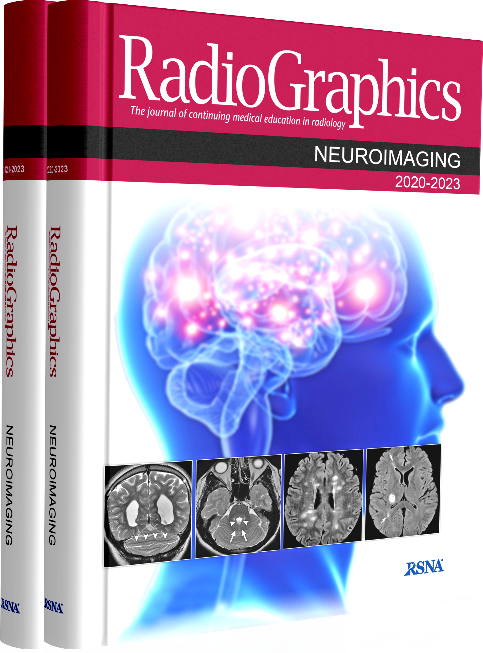
RadioGraphics یک مجله ادواری است که تحت نظارت هیئت مدیره انجمن رادیولوژی آمریکا، شرکت (RSNA) منتشر می شود، انجمن رادیولوژی آمریکا بر ماهیت همه مطالب انتخاب شده برای انتشار، نظارت می کند.
نظر به گستره وسیع اطلاعات در حوضه رادیولوژی و تخصصی شدن مطالعه و فعالیت رادیولوزیست ها در مراکز رادیولوژی قسمت های مختلف این مجلات به صورت تخصصی تفکیک شده و جداگانه جمع آوری و اراِئه می گردد.
در سالهای اخیر دانشگاه های کشور برای به روز بودن اطلاعات اساتید و دستیاران و متخصصین از این مجموعه ها برای امتحانات برد و کارگاه های مدون آموزشی نیز بهره می برند
امید است این مجموع ها که که به صورت تخصصی جمع آوری شده و شامل جدیدترین یافته های رادیولوژی جهان است در رشد سطح علمی متخصصین کشور راه گشا بوده باشد.
این مجموعه شامل این مقالات می باشد.
*** # Radiographics: Neuroimaging 2020-2023
Radiographics – The journal of continuing medical in radiology Neuroimaging 2020-2023
2020-2021
• Multimodality Imaging of Dementia: Clinical Importance and Role of Integrated Anatomic and Molecular Imaging
• CT Myelography: Clinical Indications and Imaging Findings
• High-Resolution Laryngeal US: Imaging Technique, Normal Anatomy, and Spectrum of Disease
• RadioGraphics Update: White Matter Diseases with Radiologic-Pathologic Correlation
• Intramedullary Masses of the Spinal Cord: Radiologic-Pathologic Correlation
• Invited Commentary: A Framework for the Differential Diagnosis of Benign and Malignant Intramedullary Tumors
• Temporal Bone Trauma: Typical CT and MRI Appearances and Important Points for Evaluation
• Mechanisms and Origins of Spinal Pain: from Molecules to Anatomy, with Diagnostic Clues and Imaging Findings
• Parathyroid 4D CT: What the Surgeon Wants to Know
• Understanding Pediatric Neuroimmune Disorder Conflicts: A Neuroradiologic Approach in the Molecular Era
• Anatomy, Imaging, and Pathologic Conditions of the Brachial Plexus
• Nonepithelial Tumors of the Larynx: Single-Institution 13-Year Review with Radiologic Pathologic Correlation
• Diagnosis of Skull Base Osteomyelitis
• Imaging of Malignant Minor Salivary Gland Tumors of the Head and Neck
• Differential Diagnosis of Facet Joint Disorders
• A Practical Approach to Diagnosis of Spinal Dysraphism
• Postoperative Imaging of the Temporal Bone
• Differential Diagnosis of Corpus Callosum Lesions: Beyond the Typical Butterfly Pattern
• Imaging Spectrum of Calvarial Abnormalities
• Practical Approach to Radiopaque Jaw Lesions
• Invited Commentary: Key Concepts of CT for Penetrating Abdominopelvic Injuries
• Primary Tumors of the Pituitary Gland: Radiologic-Pathologic Correlation
• MR Angiography Series: Neurovascular MR Angiography
2022
• Central Nervous System Systemic Lupus Erythematosus: Pathophysiologic, Clinical, and Imaging Features
• Neuroradiology in Transgender Care: Facial Feminization, Laryngeal Surgery, and Beyond
• Invited Commentary: Advancing the Field of Transgender Radiology
• Normal Variants of the Oral and Maxillofacial Region: Mimics and Pitfalls
• Imaging the External Ear: Practical Approach to Normal and Pathologic Conditions
• Imaging Intracranial Aneurysms in the Endovascular Era: Surveillance and Posttreatment Follow-up
• Invited Commentary: Maintaining Radiologist Relevance in the Endovascular Era of Cerebrovascular Aneurysm Treatment
• Adverse Radiation Therapy Effects in the Treatment of Head and Neck Tumors
• Imaging Aspects of the Hippocampus
• Quantitative Susceptibility Mapping: Basic Methods and Clinical Applications
• Anatomy and Diseases of the Greater Wings of the Sphenoid Bone
• Major Changes in 2021 World Health Organization Classification of Central Nervous System Tumors
• Deadly Fungi: Invasive Fungal Rhinosinusitis in the Head and Neck
2023
• Arterial Spin Labeling: Techniques, Clinical Applications, and
• Interpretation
• Imaging the Cerebral Veins in Pediatric Patients: Beyond Dural Venous Sinus Thrombosis
• Imaging Approach for Cervical Lymph Node Metastases from Unknown Primary Tumor
• Cochlear Implantation: Systematic Approach to Preoperative Radiologic Evaluation
• Systematic Approach to Pediatric Macrocephaly
• Focused Abbreviated Survey MRI Protocols for Brain and Spine Imaging
• Congratulations to the 2023 Editorial Fellows
• Radiologist’s Role in Anti-Amyloid Therapy for Alzheimer Disease
• Dural and Leptomeningeal Diseases: Anatomy, Causes, and Neuroimaging Findings


